
Femoral Derotation Osteotomy
If your knees point inward when you walk ("pigeon-toed") or outward ("duck-footed"), the underlying cause may be abnormal rotation of your thigh bone (femur). A femoral derotation osteotomy is a surgical procedure that corrects this alignment, helping the hip, knee and foot work together more efficiently. For many patients, correcting femoral rotation can reduce pain, improve walking and running, increase confidence in movement, and help prevent ongoing joint problems.
Dr David Agolley treats abnormal femoral rotation and performs femoral derotation osteotomy on the Gold Coast. To book a consultation with Dr Agolley to learn more about your options, do not hesitate to
contact us at
Dr Agolley's Tugun Clinic or
Southport Clinic.
Understanding the Symptoms of Abnormal Femoral Rotation & Femoral Version
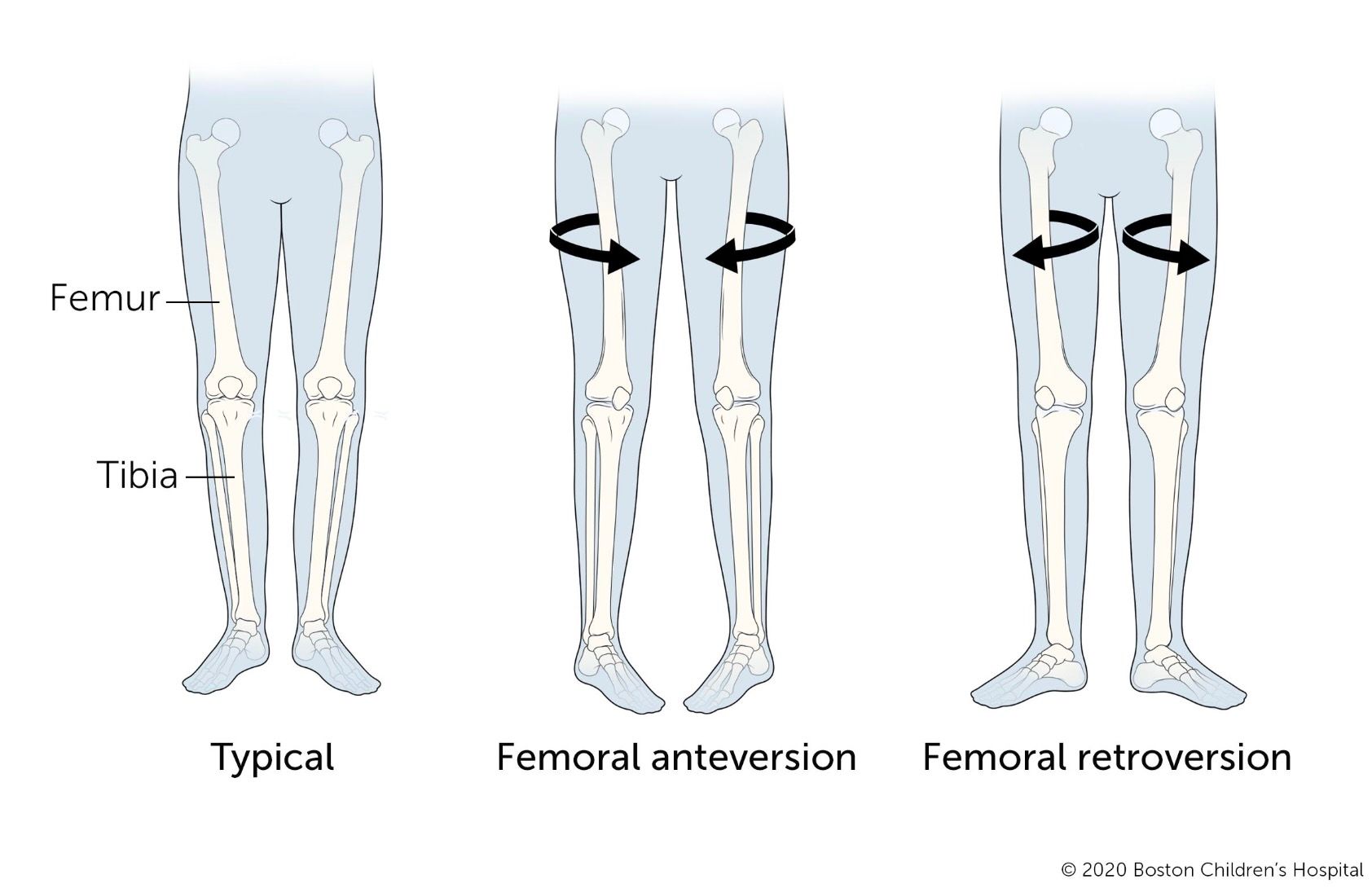
The femur naturally has a small amount of rotation. When that rotation is excessive, it can change how the hips and knees move.
Femoral Anteversion
The knees and feet tend to point inward ("pigeon-toed"), which may contribute to hip pain, kneecap instability and tripping.
Femoral Retroversion
The knees and feet tend to point outward ("duck-footed"), which may contribute to hip impingement, altered gait and difficulty running.
Abnormal Femoral Version
Research shows that abnormal femoral version can significantly affect hip stability, kneecap tracking, and overall lower-limb function. Abnormal femoral version can present differently depending on whether the femur is excessively anteverted or retroverted, but common symptoms include:
- Anterior knee pain, particularly around or behind the kneecap
- A feeling of the kneecap catching, shifting or dislocating, especially with pivoting sports
- Hip pain, catching or a pinching sensation with deep hip flexion, consistent with femoroacetabular impingement
- Awkward, inefficient or fatiguing gait, particularly noticeable during sport
- Compensatory foot or lower back symptoms from years of altered lower limb alignment
Because these symptoms overlap with several other hip and knee conditions, a proper rotational assessment is important before attributing them to femoral version.
What is a Femoral Derotation Osteotomy?
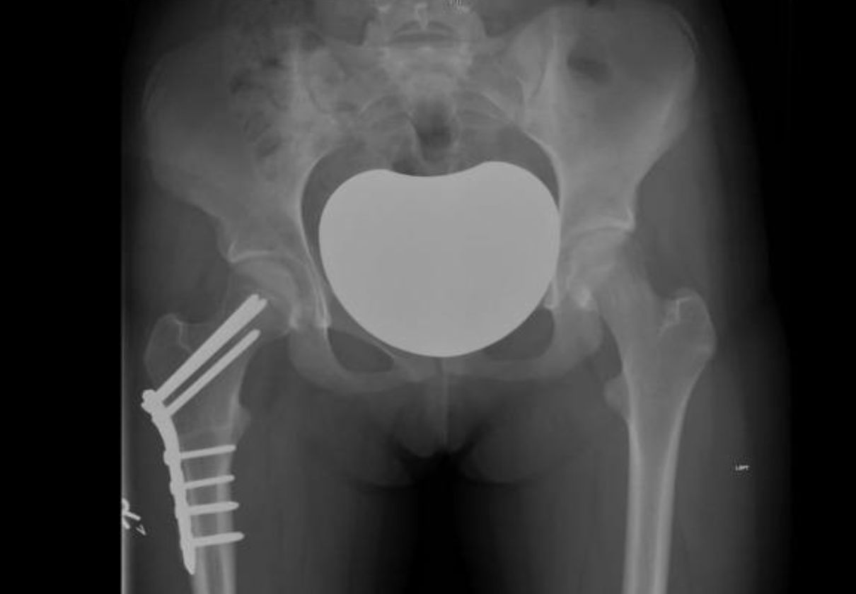
A femoral derotation osteotomy is an operation that rotates the femur into a more normal position. During surgery, the thigh bone is carefully cut, rotated to the planned angle, and held securely with either:
- a plate and screws, or
- an intramedullary rod with locking screws.
As the bone heals, it joins together in its new alignment, improving the mechanics of the entire leg.
Unlike a hip replacement, this procedure preserves your natural hip joint while correcting the underlying bone alignment.
How is Femoral Version Diagnosed?
Accurate diagnosis is essential before considering surgery.
Your assessment would typically include:
- A detailed clinical examination
- Gait assessment
- Measurement of hip rotation
- CT scanning to precisely measure femoral version
CT imaging remains the most accurate method for measuring femoral rotation and planning surgery.
Who May Benefit from a Femoral Derotation Osteotomy?
Surgery is usually considered when symptoms persist despite non-operative treatment.
You may benefit if you have:
- Persistent hip, knee or leg pain related to abnormal femoral rotation
- In-toeing or out-toeing that causes tripping, fatigue or difficulty walking
- Kneecap instability or patellofemoral maltracking
- Hip impingement symptoms or labral tears related to abnormal femoral version
- Difficulty participating in sport because of poor leg alignment
- Symptoms that have not improved with physiotherapy, strengthening or bracing
How is a Femoral Derotation Osteotomy operation performed?
The procedure is carefully planned before surgery to achieve the correct amount of correction.
Step 1 – Anaesthetic
You'll receive a general anaesthetic so you're completely asleep during the operation.
Step 2 – Incision
An incision is made on the outside of the thigh.
Step 3 – Osteotomy
The femur is carefully divided.
Step 4 – Rotation
The bone is rotated into the planned position using pre-operative planning and intra-operative checks.
Step 5 – Fixation
The corrected position is secured using either:
- a plate and screws, or
- an intramedullary rod with locking screws.
Step 6 – Closure
The muscles and tissues are repaired and the skin is closed with sutures or staples.
Recovery After Femoral Derotation Osteotomy Surgery
Recovery is gradual as the bone heals and the muscles adapt to their new alignment.
Physiotherapy plays an important role in restoring strength, balance and confidence after surgery.
Most patients notice that their walking pattern feels straighter relatively early, although strength and endurance continue improving over several months.
Typical Recovery Timeline
| Stage | What To Expect |
|---|---|
| Hospital stay | 1–2 nights |
| Walking with crutches | 6–8 weeks |
| Return to driving (left leg) | Around 4 weeks |
| Return to driving (right leg) | Around 4 weeks |
| Office work | 2–4 weeks |
| Gentle sport | 3–6 months |
| Hardware removal (if required) | Around 12 months |
Risks of Femoral Derotation Osteotomy Surgery
As with any major bone surgery, complications can occur.
Potential risks include:
- Infection
- Bleeding
- Blood clots (DVT)
- Nerve or blood vessel injury (rare)
- Delayed bone healing or non-union
- Under- or over-correction of rotation
- Hardware irritation
- Stiffness
- Persistent pain
- Anaesthetic complications
Careful surgical planning and rehabilitation help minimise these risks.
Expected Outcomes of Femoral Derotation Osteotomy Surgery
Studies have shown that appropriately selected patients generally experience:
- Improved walking pattern
- Better hip and knee mechanics
- Reduced pain
- Improved kneecap stability
- Increased ability to participate in sport and daily activities
The best results occur when surgery is combined with a structured rehabilitation program.
Femoral Derotation Osteotomy for Children
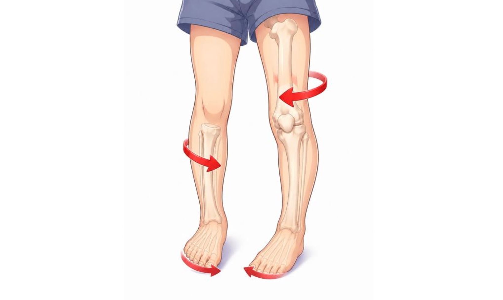
Some children develop excessive inward rotation of the femur that causes their legs to turn inward while walking.
When the problem persists and causes functional difficulties, a femoral derotation osteotomy may help make walking easier and more comfortable.
Before Your Child's Surgery
Your child's treatment team will advise whether additional tests or scans are needed before surgery.
You may also meet with:
- a physiotherapist
- an occupational therapist.
Before surgery, the hospital will contact you with:
- arrival time
- fasting instructions
- expected hospital stay.
It's also helpful to:
- inform your child's school
- organise time away from work
- arrange family support during recovery.
During the Operation
Your child will receive a general anaesthetic.
The procedure involves:
- cutting the upper part of the thigh bone
- rotating the bone so the knee faces forward
- securing it with plates and screws.
If necessary, both legs can be treated, although this depends on individual circumstances.
After Your Childs Surgery
Pain relief is carefully managed using a combination of:
- nerve block
- intravenous medication
- oral pain medicine.
Your child's surgeon will advise when they can:
- go home
- begin putting weight through the leg
- stop wearing a brace (if one is required).
Recovery varies depending on bone healing and whether other procedures were performed at the same time.
Femoral Derotation Osteotomy FAQs
How do you know if my femur is rotated abnormally?
We combine a clinical examination, gait assessment and CT scanning to accurately measure femoral version and determine whether abnormal rotation is contributing to your symptoms.
Can both legs be operated on?
Yes. If both femurs are abnormally rotated, surgery can be performed, although it is usually done one side at a time.
Will the plate or rod need to be removed?
Not always. Some implants remain permanently, but many patients have them removed after the bone has fully healed, typically around 12 months after surgery.
Is this the same as a hip replacement?
No. This operation preserves your natural hip by correcting the bone alignment rather than replacing the joint.
What is the success rate?
Most patients experience meaningful improvements in walking, pain and hip and knee function when surgery is appropriately planned and followed by rehabilitation.
How soon will I notice improvement?
Many patients notice straighter walking soon after surgery, while strength, confidence and overall function continue improving over several months.
Ready to Learn More About Correcting Pigeon-toe & Out-toeing Conditions?
If hip pain, knee pain or an abnormal walking pattern is limiting your life, I'd be happy to assess your symptoms, review your imaging and discuss whether a femoral derotation osteotomy is the right solution for you.
Together, we'll create a treatment plan tailored to your lifestyle, sporting goals and long-term joint health.
Book a Consultation
Discuss your symptoms, review your imaging and find out whether femoral derotation osteotomy is right for you.
References
1. Nelitz M et al. Femoral Derotational Osteotomies. Current Reviews in Musculoskeletal Medicine. 2018.
Open-access article
.
2. Orthopaedic Specialty Institute. Femoral Derotational Osteotomy Patient Information. Used for general patient education concepts and procedural descriptions.
3. Femoral version diagram by Boston Childrens Hospital.